|
|

| Services
home
> Technical Support >
|
Vascular Imager
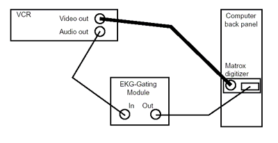
| 1. Cable Connection using
the Gating |

| 2. Matrox Drivers and Installation |
There are two separate sets of Matrox drivers - choose carefully based on your Imager version:
Contact MIA for support.
| 3. Common Problems and
Solutions - Imager |
-
After starting Vascular Imager, the following error message
is shown:
Solution: Only one Vascular Imager can
be invoked at any given time. Because there is only one
frame grabber board inside the computer, so it can serve
only one instance of the Imager application.
-
Acquisition time or number of frames cannot be entered -
a smaller number appears instead.
Solution: The length of sequence exceeds
the allowed limits or the memory space available. Decrease
the length of the acquired image sequence to be below the
limits specified in Section 11.2.3 in the User's Manual.
If the numbers are below these limits, and other applications
are running, stop the other applications, and re-enter the
numbers again.
-
The Imager appears to be hanging in the Trigger mode.
Solution: In the trigger mode, the video
display is updated ONLY upon the arrival of a trigger signal
- even the display is control by the trigger signal. If
there is no incoming trigger signal, the imager is not hanging
but rather waiting for the trigger signal to come. To trouble
shoot the lack of trigger signal (cabling problem, VCR problem,
etc.), you may switch the acquisition mode, from Triggered
back to the Timed mode to have the regular video display
and check on the trigger signal path.
-
EKG-gated triggering is too frequent and acquires image frames
between R-waves – too frequent triggering.
Solution: Triggering signal is incorrect.
This may be caused by the signal itself (contact your EKG
system technician) or by the incorrect (too high) voltage
level of the triggering signal at the digitization board
triggering input. In the latter case, adjust (decrease)
the gain of the hardware Vascular EKG Gating Module to achieve
correct triggering. See also Chapter 12 in the User's Manual.
-
EKG-gated triggering is not frequent enough and triggered
image acquisition skips cardiac cycles – too infrequent
triggering.
Solution: Triggering signal is incorrect.
This may be caused by the signal itself (contact your EKG
system technician) or by the incorrect (too low) voltage
level of the triggering signal at the digitization boar
triggering input. In the latter case, adjust (increase)
the gain of the hardware Vascular EKG Gating Module to achieve
correct triggering. See also Chapter 12 in the User's Manual.
-
Vascular EKG Gating Module does not provide correct gating
data while it functioned properly in the past.
Solution: Check the battery of the Vascular
EKG Gating Module (red control light on) and replace if
necessary. Adjust the gain otherwise. See also Chapter 12
in the User's Manual.
-
Vascular Imager is not responding after clicking "Save"
in the saving window and before the study information window
pops up.
Solution: This is normal, the application
is not crashing. Saving the acquired image data takes time.
The length is dependent on the computer speed and the number
of frames acquired.
-
Vascular Imager sometimes requires more time than specified
to acquire an image frame.
Solution: Image acquisition is a very
demanding application. To maximize the performance, do not
run any other Windows application during image acquisition.
The computer used for image acquisition should not be connected
to any network during frame grabbing. See Section 11.3.1
in the User's Manual.
-
Vascular Imager does not let me save long image sequences
despite the fact that I have sufficient amount of RAM memory
installed.
Solution: Please, check your operating
system. You must be running Windows NT, 2000, or XP to be
able to handle large-size reliably. Windows 95/98?Me operating
system was not designed to deal with files that are larger
than several tens of megabytes. If you are running Windows
95, 98 or Me, upgrade to Windows NT, Windows 2000 or Windows
XP.
-
When using triggered acquisition, some triggers are missed.
Solution: Make sure the duplicate-triggering
option is configured correctly. See Section 11.3.8 in the User's
Manual.
- Sometimes while exiting the Vascular Imager application, there may be error messages displayed. The error messages are entitled MIL DLL Message or Native MIL Error.
Solution: Those error messages can be safely ignored, by clicking on the Cancel or No button on the pop-up. The errors do not affect the behavior of the Vascular Imager or its image acquisition. The errors are coming from the hardware frame grabber board due to the hand-shaking of the exiting.
|
|
| |
|
|
Vascular - clinical use
Vascular
Tools 5- FDA Approved - clinical assesment of FMD/endothelial
function of brachial artery and IMT/intimal-medial thickness of carotid
artery from ultrasound image data.
|
|
Vascular - research use
Vascular
Research Tools 6 - Vascular
Tools plus additional modules for carotid plaque analysis, vascular
compliance. Includes additional reporting flexibility, well suited
for clinical trials. Available as software or as turn-key integrated
workstations, for investigational use only. |
|
Aorta - research use
Aortic Analyzer
... software for quantitative analysis of aortic morphology and function for MR or CT data in 3D and 4D
|
|
Cartilage -
CartA
... software for quantitative analysis of cartilage MR image data
in 2D and 3D
|
|
|


