
|
|

| Services
home
> Technical Support >
|
Brachial Analyzer
and Brachial Analyzer for Research.
| Common Problems and Solutions |
-
The analysis is slower than usually when the Results window
is open and the calculated diameter function is displayed
in real time.
Solution: Real-time display of the brachial
diameter and the frame status symbols (*/r/e/i/e) is useful
for analysis process monitoring but computationally demanding.
The slow-down should not be noticeable on a faster computers.
On slower machines, close the Results window when performing
the sequence analysis.
-
In image sequences with pronounced intimal layer at the far
border, the first stage training correctly identifies the
desired border (e.g., the medial layer) but the second stage
border detection identifies a different well-visible border
of the far wall.
Solution: Train in another image frame
with visually better image quality.
-
The region of interest is not displayed.
Solution: Make sure that the ROI display
button  is selected (pressed). This is an on-off toggle. is selected (pressed). This is an on-off toggle.
-
The Brachial Analyzer does not show the resulting borders.
Solution: Please make sure that the border
display button  is selected (pressed). This is an on-off toggle. is selected (pressed). This is an on-off toggle.
-
The Brachial Analyzer sometimes does not show the resulting
borders, the ROI box, and/or the calibration markers.
Solution: This may be caused by an unfortunate
selection of colors used to display borders and lines. To
check that, click "Options" from the "View"
menu, make sure the "Display Colors, Line Styles"
tab is selected and make sure the selected colors (shown
on the big squares) are visible. Bright colors are preferred
for both the ROI and the border. In other words if a black
color is selected you will have trouble seeing the border
or ROI on an image. Make changes if necessary and click
"OK" or "Apply".
-
Some AVI image data can not be opened.
Solution: Higher level of compression is not supported in order to achieve good quality of the analysis. Adjust the application that generates the AVI image data to use the non-compressed or minimal-compressed option for the image data.
-
Unable to reset ROI.
Solution: Open the “Initialization” control panel (Action -> Initialize) and go to Step 1 (you may or may not need to click the “Start Over” button, based on the status of your current analysis):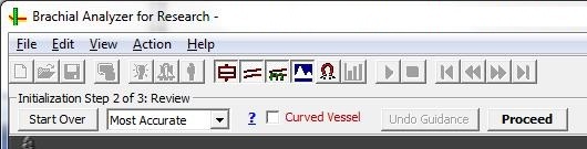
This screen appears: 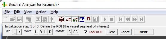
Click the “Clear” button, then click in the middle of the vessel. A good and orderly ROI will appear. Proceed normally.
|
| |
|
| |
|
|
Vascular - clinical use
Vascular
Tools 5- FDA Approved - clinical assesment of FMD/endothelial
function of brachial artery and IMT/intimal-medial thickness of carotid
artery from ultrasound image data.
|
|
Vascular - research use
Vascular
Research Tools 6 - Vascular
Tools plus additional modules for carotid plaque analysis, vascular
compliance. Includes additional reporting flexibility, well suited
for clinical trials. Available as software or as turn-key integrated
workstations, for investigational use only. |
|
Aorta - research use
Aortic Analyzer
... software for quantitative analysis of aortic morphology and function for MR or CT data in 3D and 4D
|
|
Cartilage -
CartA
... software for quantitative analysis of cartilage MR image data
in 2D and 3D
|
|
|